YOUR FAVORITE PT
  • Home
  • About
  • Blog
  • Case Studies
  • Events
  • Services
  • Contact
  • Feedback

Case Studies: Is Your Clinical Reasoning Within Reason?

  • Do you ever catch yourself falling into the habit of prescribing the same group of exercises for every patient/client presenting to PT with knee pain?

  • When your patients arrive to your office with intense leg pain that radiates from their gluteal region to their great toe, do your methods to differential diagnosis the root cause of their symptoms get the patient better in 1-2 visits?

  • Do you feel like special tests make your clinical exam even more confusing at times and carry more weight in your clinical decision making than they should?

  • How important is it to you to establish a physical therapy diagnosis?

  • Are you comfortable with carrying out a plan of care based solely on a medical diagnosis?


Our profession has shifted vastly over the years in which the days of being a mere technician are long gone. Our patients are expecting more from us and our presence within the medical community has become more prominent as evidence-based practice has evolved. Our goal as orthopedic specialists is to stay up to date on best practice in the realm of orthopedic physical therapy. This also involves critically appraising current research and being able to implement advanced clinical reasoning. The more foundational knowledge you're equipped with, the better you help your patients.  Let's approach our patients with humility and active listening, remembering they've come to us during a moment of vulnerability seeking help. 


"The orthopedic manual therapy examination consists of two parts, a differential diagnostic examination and a bio-mechanical examination" - Jim Meadows


The main objective of the case studies listed below is to primarily address the differential diagnostic examination, not give medical advice.
Case: The 14-Year-Old Diver

7/2/2021

2 Comments

 
Part 1: The patient is a 14 year old female presenting to outpatient physical therapy with complaints of low back pain of recent onset 2 weeks ago. She denies any mechanism of injury. Her back pain is localized to the lower lumbar spine and hurts with walking, sitting for extended periods of time, bending forward/backwards as well as twisting. She describes her pain as achy at rest and sharp at times with movement. She denies any feelings of weakness, radicular pain, paresthesias or bowl/bladder changes.

Her father adds that she injured her left ankle 2 years ago during a diving incident while attempting to jump from the diving board. He mentions his daughter suffered a type 2 talus fracture requiring surgical fixation and subsequent removal of hardware 6 months later. She attempted to return to diving 8 weeks from the date of her surgery after being cleared by her surgeon. This attempt was unsuccessful due to moderate pain and weakness. She was then treated in physical therapy for approximately 1 year, but chose not return to diving. She does participate in high school cheerleading for the football team as a base and is currently 4 weeks into the season.

She is 5 feet, 2 inches tall and weighs 115 lbs.

F/u Questions for you
1. Are there any red flags? yellow flags? sinister pathology you would like to rule out?
2. Based on the information provided, what additional subjective information would you like to know and why?
3. Could you come up with at least 2 preliminary diagnoses?
4. What subjective outcome measures would you select that may provide useful information from this patient?

Part 1: summary
1. Are there any red flags? yellow flags?
- Red Flags: Any information that help decrease the chances of serious pathology such as a space occupying lesion, infectious disease or Spondyloarthropathy. Some information was provided that would help increase our confidence in there being no red flags such as no bowel/bladder changes. Though for a teenager, the wording of these questions may need to be more specific to get a more accurate response. Additional information that help with the other red flags include: 1) any recent sicknesses 2) feelings of stiffness at any point in the day and 3) pain in other joints.
- Yellow Flags: any recent growth spurts and by how much, changes in weight, changes in coordination that accompanied the most recent growth spurt, a history of scoliosis, hyper-mobility, congenital anomalies (ie., lumbarization, sacralization, can be structural or functional)
- In this case, there were no red or yellow flags

2. Based on the information provided, what additional subjective information would you like to know and why?
- Has she had any imaging performed? Yes, x-rays were negative for significant pathology. Views used were anterior/posterior and lateral. Would you want any additional imaging at this point? If so, what type of imaging and why?
- Has she had a Risser score for stages of growth or told anything about growth? per chart review, she was classified as a grade 3, indicating a slowing of growth. She was told that she is done growing for the most part.
- Why hasn’t she returned to diving? In this case she states she is nervous about returning to the diving board due to the mental imagery surrounding her original injury and that she does not feel she can trust her ankle. Her father felt he was doing the right thing by returning her to diving at 8 weeks once she was cleared by the surgeon, even though she explained she did not feel ready. 
- How did her rehab for the ankle injury go? It was slow, she ended up having quite a bit of pain from the screws before getting them removed. The surgeon also explained she had excessive scar tissue that needed to be removed.  

3. Could you come up with at least 2 preliminary diagnoses?
- spondylolysis
- spondylolisthesis
- lumbar strain
- complex regional pain syndrome (reflex neuro-vascular dystrophy)
- sacroiliac dysfunction
- mechanical causes of low back pain

4. What subjective outcome measures would you most likely provide that may provide useful information for this patient?
- Micheli Functional Scale
- Numeric Rating Scale-11 for pain  or Visual Analog Scale
- Patient Specific Functional Scale

Part 2
After digging deeper with the history, you feel confident that the patient is at a low risk of having any red flags such as neurological symptoms indicative of a spinal tumor. You do note that she has grown 3 inches in the past 6 months and has gained 30 lbs since her ankle injury. She feels she is not as flexible as she was prior to her growth spurt.  She shares with you that she has been a cheerleader for 8 years and has no previous history of back pain. Her pain is described as 2/10 at rest during your evaluation and 7/10 at worst. Her pain with trunk flexion typically occurs at end range when she attempts to return to a standing position. Her pain with trunk extension is described as sharp and sudden at times.

Objective Examination
Static standing posture:
- sway back with increased thoracic kyphosis
- no significant signs of muscle imbalance or scoliosis while in upright position
- hypertrophic erector spinae noted bilaterally

Gait: unremarkable with the exception of decreased arm swing noted bilaterally and a decreased cadence.

Assessment of spinal mobility in the cardinal planes reveals demonstrating full range trunk flexion followed by a curve reversal and onset of pain when returning to an upright position. No signs of scoliosis were noted in a fully trunk flexed position. Lateral trunk flexion is unremarkable for pain and range of motion deficits. Extension is limited early in the movement, painful and described as occurring bilaterally in her lower lumbar region. Upon observation, you also notice her willingness to move into trunk extension is diminished. Over pressure was applied to all cardinal plane movements and reproduction of pain was noted with extension only.

Neurological assessment was unremarkable with regards to strength testing of key muscles , sensation and deep tendon reflexes.

f/u Questions to part 2
1. Are you missing any important objective information? If so, what else would you like to know?
2. If you could only pick to perform 3 additional special tests, which would you choose and why?
3. Did the additional information strengthen or weaken any of your initial hypotheses?

2 Comments

Case: 23-year old with Parsonage-Turner syndrome

7/2/2021

0 Comments

 
Subjective Exam:
Your patient is a 23 year old, left hand dominant, male presenting to outpatient PT with complaints of right scapular pain that began approximately 1 year ago of insidious onset. He was diagnosed with Parsonage-Turner Syndrome by a neurologist after seeking treatment for his symptoms. Thus far, he has consulted with his primary care physician, a cardiologist, a neurologist and a physical therapist.  He denies any trauma, but does mention having a history of heavy power-lifting.  He states the pain in his right scapular region began as a pinching/burning sensation that was accompanied by a unilateral headache in the sub-occipital region lasting less than 1 hour. The onset of his symptoms typically occurred after prolonged sitting at his desk while studying. His headache only occurred during the first 2-3 days of his initial onset of symptoms.


He reports previous PT was helpful with pain through treatment that included massage, stretching, strengthening, and a TENS unit. He also describes sitting in positions with his back supported reduces his pain.  He mentions coughing, sneezing, and burping as factors that increase his symptoms. He recently started a "lifestyle" change and has since lost 20-30 pounds after becoming less active and gaining 60 lbs. He denies any changes in bladder, speech, vision, hearing, and or dizziness.

EMG results revealed chronic right suprascapular and dorsal scapular neuropathies, no other significant findings noted

Chen, S., et al. (2016). "Electrodiagnostic reference values for upper and lower limb nerve conduction studies in adult populations." Muscle Nerve 54(3): 371-377.

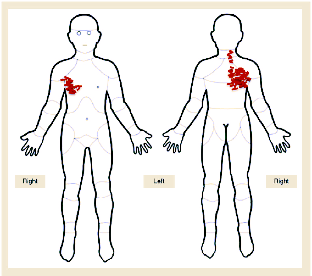
Pain chart
Picture
Observation
Height: 5'8'' Weight: 240 lbs  BMI: 36.5
Observation, Static standing posture:
- right scapula approximately 4 inches from mid-line and left scapula approximately 2 inches from mid-line
- prominent medial and inferior borders of right scapula noted upon inspection
- dowager's hump noted in upper thoracic region
- muscle atrophy noted in right posterior shoulder girdle and periscapular muscles
- normal tone, no fasciculations noted

Self Assessment?
1. Based on the history and observation, is there anything else you would like to know?
2. Do you have enough information to make a preliminary physical therapy diagnosis?
3. How does his diagnosis of Parsonage-Turner Syndrome influence his symptoms?
4. What would you include in you objective exam that may provide more insight to this case?

Objective Exam
The range of motion in his involved extremity when assessing shoulder elevation was reduced in flexion, abduction. He had approximately 150 degrees in each direction before having complaints of posterior shoulder girdle pain. He demonstrated superior border dysfunction and ipsilateral cervical lateral flexion when attempting to exceed his available range. Assessment of his thoracic extension and seated trunk rotation based on the expected available range of motion was 25% to the right (reproduction of posterior shoulder girdle pain) and 50% to the left, in addition to 50% of trunk extension. He demonstrated a reduction in chest expansion with a reproduction of his posterior shoulder girdle symptoms.
Case summary
In this case, the patient was deemed appropriate for outpatient physical therapy services. Differential diagnosis for his shoulder pain consisted of referred pain from the liver and gall bladder, cervical spine, C5-C6 more specifically, costovertebral or costotransverse joint pain and rotator cuff pathology. His symptoms of headache that were present during the acute phase of his injury were thought to be of little significance to this case as symptom onset was approximately 1 year ago. 

The patient did not have risk factors or symptoms that would increase the likelihood of his pain being visceral in origin. This includes a history of liver disease, over the age of 40, a diet low in fiber, high in cholesterol or high in fats, yellow eye coloration, complaints of nausea/vomiting, or episodes of sudden intense pain not alleviated with position change. His medical diagnosis of Parsonage Turner Syndrome was not consistent with current clinical presentation of anterior chest and posterior shoulder girdle pain detailed as burning and pinching in nature. The EMG findings of neuropathy of the suprascapular nerve and dorsal scapular nerves are significant when assessing the patient's strength and movement patterns related to shoulder elevation, but are an unlikely culprit of this patient's chief complaint. However, the suprascapular nerve, a mixed nerve, has sensory distribution to the lateral aspect of the arm and functions primarily in its motor innervation of the supraspinatus and infraspinatus muscles.

Upon further investigation, his burning sensation was diffuse throughout the posterior shoulder girdle and anterior chest. These symptoms were described as beginning posterior over the scapula with a gradual progression to the anterior aspect of his thorax. His feeling of localized pinching was reproduced with deep inhalation/exhalation, ipsilateral trunk rotation and palpation proximal to the region of the costo-transverse and costo-vertebral joints. 

After a bio-mechanical exam of the upper quadrant was performed, the patient was treated for costo-vertebral and costo-transverse joint pain and muscle imbalances related to shoulder elevation.

0 Comments

Case: The 26-year-old weekend warrior

7/2/2021

2 Comments

 
The patient is a 26-year-old male presenting to PT with complaints of neck pain of traumatic onset three weeks ago. He reports falling on his head while attempting to do a handstand push-up after finishing a CrossFit workout. He describes feeling a jolt through his head and neck followed by a period of neck stiffness. He rates his initial neck pain as 4/10 that increased to 6/10 the next day on the numeric pain rating scale. He denies any use of medications for pain and has refrained from working out at CrossFit since. He is having difficulty sleeping and has begun to experience headaches. He works full time as a clinical assistant at a physician’s office where he receives phone calls and helps with intake information for patient visits. He has difficulty driving, reading, and concentrating for extended periods of time.

Imaging was performed after visiting his primary care physician and x-rays were unremarkable

 f/u Questions for you
1. Do you have enough information to determine if this patient appropriate for physical therapy?
2. Based on the information listed above, are we able to establish any hypotheses?
3. If so, what are some competing diagnoses for this patient?
4. What additional subjective information would you like to know for this case to assist you in establishing a physical therapy diagnosis?

Part 1 summary
After further investigation, the patient reveals a sense of being lightheaded and having blurred vision immediately after landing on his head. He denied any loss of consciousness, dizziness or double vision. He has returned to exercising, but has limited himself to walking on the treadmill for up to 15 minutes and only use of light free weights at the gym. He feels more tired at work and averages about 4 to 5 hours of sleep a night. His neck pain has limited his ability to drive due to turning his head so he has limited driving outside of work and getting food. He tells you he feels like his neck just needs to be manipulated. He continues to experience blurred vision and attributes these symptoms to his lack of sleep and feeling tired. He denies any history of head/neck trauma.
f/u questions for you based on summary
1. Based on this information, what subjective outcome measures would be appropriate for this patient?
2. Are referrals to a specialist needed?
3. Based on the patient’s age and mechanism of injury or would you like any additional imaging for this patient? If so what type?
4. Considering moving forward with an objective exam, what red flag information would you want to rule out and where would you start with your exam?

Objective Exam
The patient's history does not suggest any signs of an upper motor neuron lesion or a multi-level nerve root pathology. He does not have a history of hypertension, smoking, cancer, connective tissue or autoimmune disorders, or recent infections. He has self manipulated his neck in the past after falling asleep on the couch while watching television and experiencing similar symptoms.

You decide to start with a screen of his cervical spine for potential cervical artery dysfunction and upper cervical instability that could compromise vascular and neurological structures.  You determine increased laxity in the left alar ligament, with other ligamentous testing being unremarkable in regards to symptom provocation or abnormal end-feel. You examine his willingness to move and discover he has limited cervical rotation active range of motion bilaterally. He also lacks the ability to perform proper cranio-vertebral flexion. You find this information to be clinically relevant, but you are pushed for time and want to make sure you screen other systems.

You finish your physical examination with vestibular ocular motor screening and conclude that he has visual motion sensitivity with reported symptoms of headache and fogginess, scores of 4/10 and 5/10, respectively.


Being constrained due to time, you make the decision to incorporate additional objective outcome measures in the patient's second visit to be thorough. Take some time to consider what measures you feel are clinically relevant for this patient.
2 Comments

Case - Passing out and falling head first

7/2/2021

0 Comments

 
Pt is a 42 year old female presenting to PT with c/o dizziness and neck pain after suffering a head injury during a fall 4 months ago.  She reports being asleep on the couch and then "passing out" after getting up to go to her bed. She endorses a loss of consciousness, but is unsure of how long. She lives alone with her two dogs and was unable to drive. She was taken to the ER by emergency medical services and subsequently required 12 stitches for a forehead laceration. Imaging included a CT scan of the head/face which was negative. She has a past medical history of syncope as a teenager in which she had 2 separate episodes while getting out of the bed, but none up until now. She describes her previous episodes of syncope being lesser in intensity and without head injury

Current Medications:
Cyclobenzaprine (Flexeril)and women's multivitamins

1. Based on the information provided. Is this patient an appropriate physical therapy candidate? If not, what would make you feel more comfortable proceeding with your examination?
2. Are there any red flags? yellow flags? Is there any sinister pathology you would like to rule out?
3. Based on the information provided, what additional subjective information would you like to know and why?
4. Could you come up with at least 2 preliminary diagnoses?
5. What subjective outcome measures would you select that may provide useful information from this patient?

Part 2
After further examination, you discover the patient has been seen by cardiology and had an electrocardiogram performed that was unremarkable. You question the patient further about her dizziness and determine that it is not likely true vertigo. You begin to consider orthostatic hypotension as a potential diagnosis. After additional questioning, the patient tells you the cardiologist tested for and ruled out orthostatic hypotension. She denies a history of anemia. She does mention that it has been suggested by a physician in the past that she may have hypovolemia. She was given suggestions of dietary changes she could make.  When she does feel dizzy, it lasts anywhere from a few minutes to hours at a time. She describes it as occurring every day with the sensation of being on a boat.  She does get headaches occasionally in her sub-occipital region that typically are associated with neck pain. She denies blurred or double vision, heart palpitations, photophobia, memory loss, difficulty concentrating or feelings of dehydration.
f/u questions
1. Is there anything else we need to know before proceeding with our objective examination?
2. What screening tools or assessments do you plan to include in your objective examination?
3. What outcome measures do you think will prove most useful in this particular case?

Part 3
You question the patient further about her neck pain before beginning your objective examination. You discover she did not receive imaging for her neck. Mentally, you automatically go to the Canadian C-spine rules for determining the use of imaging and feels she fit the profile for radiography of her cervical spine. Based on her acuity, you become less concerned of significant injury and move on. She describes her neck pain as unilateral, on the right side primarily. Her pain is in the right suboccipital region and presents as referred pain in the form of soreness in her upper trapezius/scapular region. She denies radiating/radicular pain, paresthesia or feelings of weakness. Her neck pain is reproduced with sustained right cervical rotation and left side-bending.  She has been sleeping on stomach and says she wakes up with pain/stiffness in neck.  Motrin and massage help.  She denies other falls/clumsiness or changes in bowel/bladder habits.
Objective Exam
BP: 110/70 mmHg
Pulse: 87
Height: 5'8'' Weight: 135 lbs

Observation: flat back posture with limited thoracic kyphosis and increased cervical lordosis
Gait: unremarkable
Dermatomal testing: unremarkable
Key muscle testing of cervical nerve roots: unremarkable

Oculomotor exam: unremarkable
Vestibular Ocular Reflex Testing:
 - Head thrust (horizontal): negative bilaterally
 - Head thrust (vertical): negative bilaterally
 - Visual acuity difference: 0

Cervical AROM
- flexion: 40 deg
- extension: 30 deg with reported generalized posterior neck pain
- sidebending: 35 deg (right), 30 deg (left) with reported right side neck pain
- rotation right: 48 deg with reported right sided neck pain
- rotation left: 50 deg

Positional Testing
- Dix Hallpike: negative bilaterally
- Modified Sidelying test: negative bilaterally
- Head roll test: negative bilaterally
- Cervical torsion test: negative bilaterally

Sensory organization testing: unremarkable

Part 4
You feel you have more than enough information to move forward. You decide to assess for potential cervical impairments and in your scan of the cervical spine you find areas needing a more detailed bio-mechanical exam. It is here you discover an region of hypo-mobility with PAIVMs of the right C2/C3 facet joint and an area of  hyper-mobility at the right C5/C6 facet joint. To be thorough, you examine the thoracic spine for physiological movements and determine limited extension in the upper thoracic region.

You decide to pursue ruling out autonomic dysfunction and exercise intolerance by administering the Buffalo Concussion Treadmill test. The results are significant for exercise intolerance with a reproduction of dizziness (greater than 3 point difference on Likert scale) that stopped the test at 6 minutes.

What do you do with this information? Are her symptoms of dizziness and neck pain related?

Considering the multitude of trajectories in the management of post concussion syndrome, you have several avenues to consider in the treatment of this patient. Implementing sound clinical reasoning, you must prioritize your findings in a manner that optimizes this patient's care.
0 Comments

    Categories

    All
    Lower Quadrant
    Lumbar Spine
    Neck Pain
    Shoulder
    Upper Quadrant
    Vestibular/Concussion

  • Home
  • About
  • Blog
  • Case Studies
  • Events
  • Services
  • Contact
  • Feedback